CT Atlas
Cross-sectional CT anatomy on real scans, labelled in 3 planes. Quiz. Pay once.
Get it on Google Play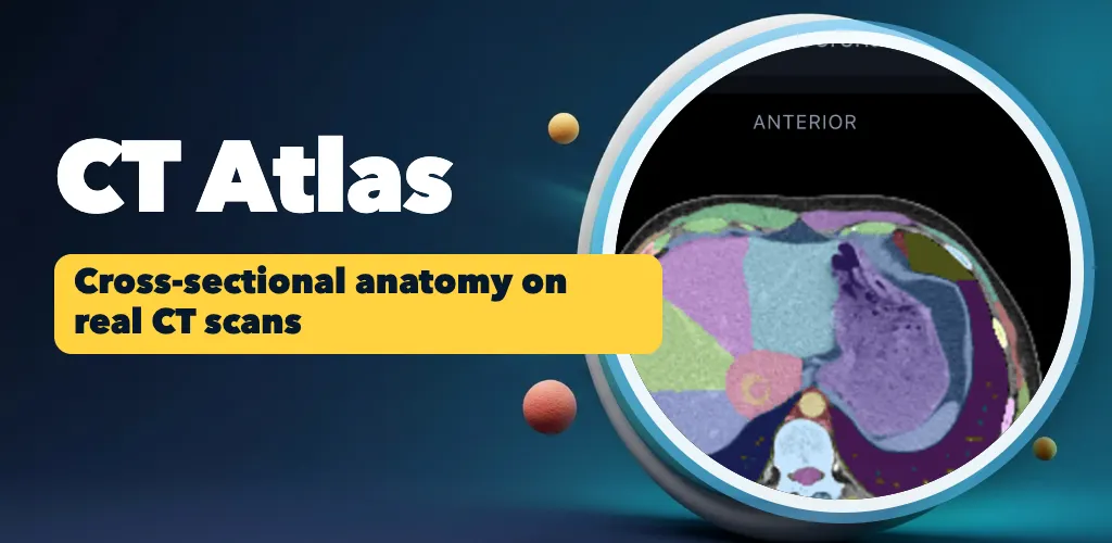
For study only. This does not replace advice or diagnosis from a healthcare professional.
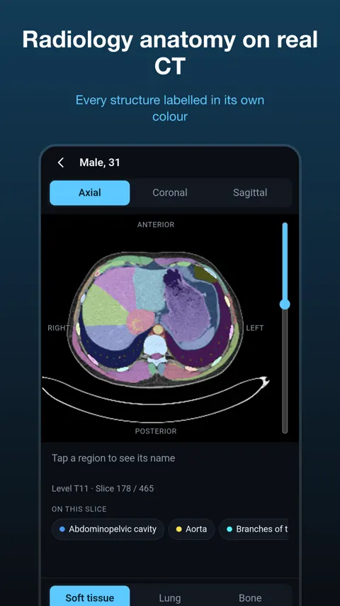
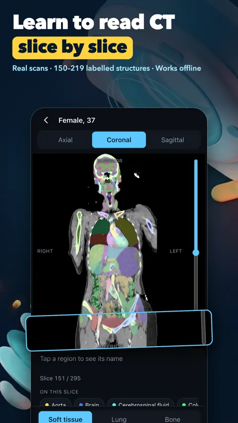
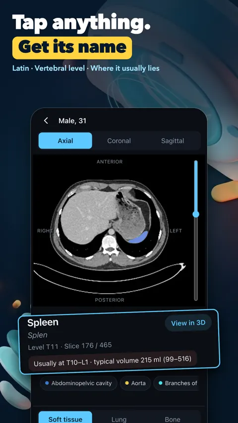
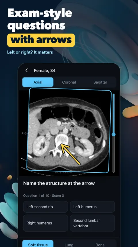
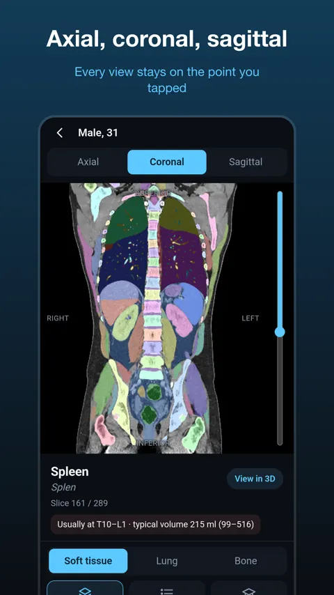
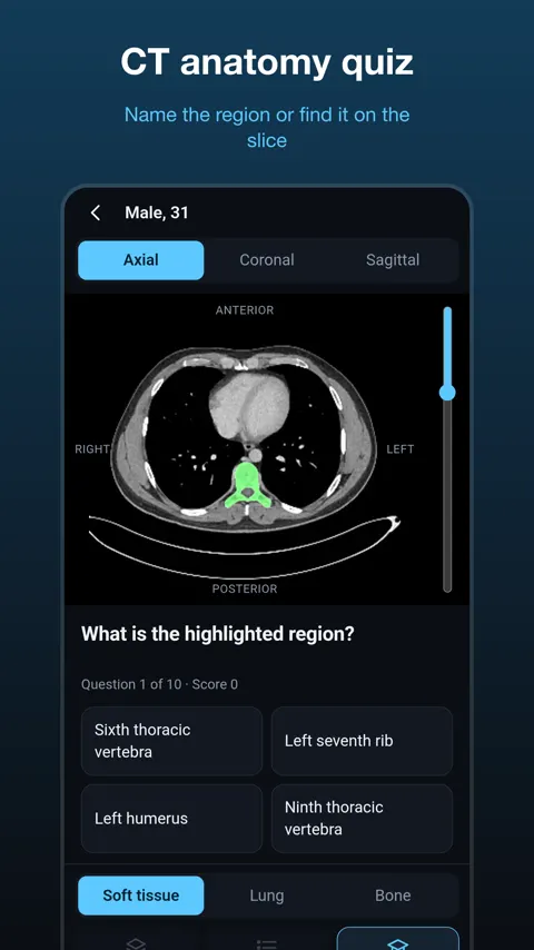
CT Atlas teaches cross-sectional anatomy on real CT scans. You scroll through slices in the axial, coronal and sagittal planes, tap any region to see its name, and then quiz yourself until the slices make sense without labels. It is made for medical students, radiology residents, radiographers, CT technologists, radiation therapists, nurses and anyone preparing for anatomy or imaging exams.
Why cross-sections are hard to learn
Most people learn anatomy from drawings that show a whole organ. A CT slice shows a flat cut through many organs at once, in grey shades, and the same structure looks different on each slice. Learning to read this takes repetition: what sits next to what, how a structure changes as you scroll, and how the three planes relate to each other. CT Atlas keeps the point you tapped in place when you switch planes, so you can follow one structure through all three views.
What is in each scan
The scans are real, not drawings: men and women, a child, a CT angiography and a whole-body scan from head to pelvis. Each scan has 150 to 219 labelled structures, including organs, bones, vessels, muscles, liver segments, heart chambers, brain and neck. Names appear in English and Latin, plus Spanish, Portuguese, French, German, Russian and Vietnamese. You can switch between soft-tissue, lung and bone windows, pinch to zoom, and search for any structure to jump to the slice where it is largest.
Knowing the vertebral level
Every axial slice shows its vertebral level. This is a useful anchor, because radiology reports and teaching often describe structures by level. For each structure you can also see the vertebral levels where it usually lies and its typical volume for the patient’s age and sex, measured on 1,119 real scans. The pancreas, for example, usually lies at T12 to L2.
Testing yourself
Quizzes are grouped by topic: organs, bones, muscles, heart and vessels, or vertebral levels. You might name a highlighted region, find a structure on the slice, or say which vertebral level an axial slice is at. Your score for each topic is kept, and the structures you miss come back first. That spacing is what turns recognition into recall.
A study plan that works with the app
Start with one region. Scroll the axial slices with labels on, then try the quiz without looking at the labels. Switch to coronal and sagittal to see the same organs from another side. Return to the vertebral level questions last, because they build on knowing the organs.
CT Atlas is a study tool for normal anatomy. It works without a network once scans are downloaded, and has no account, no sign-in and no ads.
Can I open my own CT scans in the app?
No. CT Atlas is an educational atlas of normal anatomy. It does not open your own scans and is not a medical device.
Where do the scans come from?
The images come from the TotalSegmentator CT dataset, licensed CC BY 4.0. Full credits are in the app.
Which languages are structure names in?
English and Latin (Terminologia Anatomica), plus Spanish, Portuguese, French, German, Russian and Vietnamese.
Does it work offline?
Yes. Scans are downloaded once and kept on your device, so you can study without a network.